
www.medical-devices.tech
21
'26
Written on Modified on
Mobile C-Arm Fluoroscopy System Architecture for Intraoperative Surgical Guidance
Siemens Healthineers introduces the Cios Go ID mobile C-arm with integrated display to streamline surgical imaging workflows in constrained environments.
www.siemens-healthineers.com
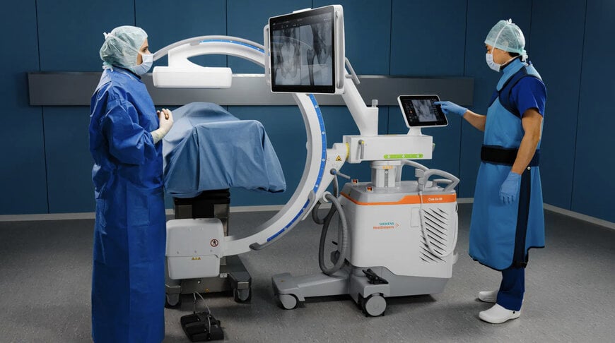
In high-turnover surgical suites, trauma bays, and outpatient surgery centers, floor space is strictly constrained by anesthesia stations, surgical tables, and sterile field perimeters. Conventional mobile fluoroscopy systems typically depend on a separate, dual-monitor trolley connected via umbilical cabling, creating cable clutter, trip hazards, and positioning bottlenecks. To resolve these spatial and workflow constraints, medical imaging manufacturer Siemens Healthineers introduced the Cios Go mobile C-arm platform, featuring the Cios Go ID variant with an onboard integrated display designed to deliver full intraoperative imaging capability on a unified mobile chassis.
Intraoperative Workflow Optimization and System Architecture
The system was showcased at the German Congress of Orthopedics and Traumatology (DKOU 2026). The Cios Go ID is engineered to support routine surgical interventions across orthopedic, trauma, spine, vascular, and minimally invasive procedures. In these environments, procedural efficiency directly correlates with clinical throughput and operational cost control.
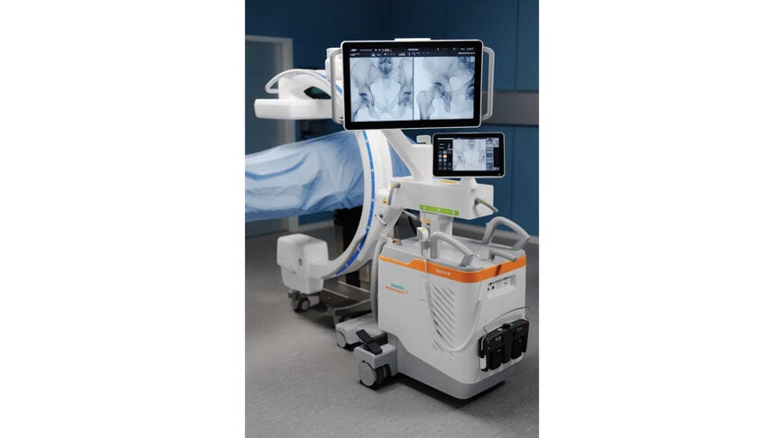
The system combines the X-ray generator, articulated C-arm assembly, flat-panel detector, and a high-resolution 4K touchscreen monitor onto a single wheeled base. Eliminating the external monitor cart reduces the total physical footprint within the sterile field, allowing surgical personnel to position the viewing display directly along the surgeon's line of sight without repositioning secondary equipment. For facility transit, an integrated monitor arm lock secures the articulated display, and an onboard standby mode allows technical staff to unplug the device, navigate corridors between surgical suites or emergency rooms, and restore full operational state upon reconnecting without a complete system reboot.
Imaging Hardware and Radiological Operating Mechanisms
At the core of the Cios Go platform is the proprietary Retina Imaging Chain, configured to uphold the radiological safety principle of ALARA (As Low As Reasonably Achievable) by optimizing radiation dose efficiency while maintaining bone and soft-tissue contrast.
The platform supports monoblock high-frequency X-ray generators configured at either 3.5 kW or 5 kW electrical power ratings. Radiation capture is handled by solid-state flat-panel detectors available in active imaging areas of 20 cm by 20 cm for extremity and targeted orthopedic interventions, or 31 cm by 31 cm for broader anatomical coverage in pelvic, spine, and vascular applications.
Digital image processing routines are automated through intelligent software functions:
- SpotAdapt: Analyzes localized anatomical density within an operator-defined region of interest, automatically rebalancing exposure parameters, brightness, and contrast curves to prevent image blooming around dense hardware implants or underexposure of adjacent bony trabeculae.
- Live Zoom: Allows clinicians to magnify targeted zones using touchscreen gesture controls directly at the table side, preserving spatial resolution for assessing micro-fractures and hardware alignment.
- Laser Guidance and Digital Targeting: A green laser-light localizer projects onto the patient surface for radiation-free beam alignment, while a Target Pointer overlays spatial reference trajectories on the live image to assist in the axial guidance of Kirschner wires (K-wires) and cannulated pedicle screws.
- Live Graphical Overlay: Permits clinicians to execute freehand anatomical annotations and trajectory sketches directly across the touch interface.
Clinical Usability Across Multidisciplinary Settings
Minimally invasive operations depend heavily on fluoroscopic verification to assess joint reduction, implant seating, and mechanical alignment without extensive anatomical exposure. User interface complexity often requires dedicated radiologic technologists to navigate multi-step submenus during surgery. The Cios Go architecture utilizes guided exam protocols and multi-touch operational profiles to allow rapid configuration with minimal orientation time across diverse clinical departments.
"Readiness is essential in our everyday work. We do not have time to keep operating rooms waiting, because every second in surgery comes at a high cost," said Niclas Riege, technologist at the Department of Trauma Surgery and Orthopedic Surgery, University Hospital Erlangen. "Cios Go ID combines a compact design with modern touch technology. The integrated display is easy to position according to where the surgeon needs to look."
The clinical versatility of the single-chassis footprint in variable facility architectures was affirmed by surgical specialists involved in initial evaluations.
"With its small footprint, Cios Go ID can fit not only in a small OR, but also in the emergency department or an outpatient setting," said Johannes M.M. Groh, MD, Consultant Trauma and Orthopedic Surgeon at University Hospital Erlangen. "The integrated monitor can be positioned exactly where it is needed, providing a clear view even in complex procedures involving multiple surgeons."
Additional Context:
This section details technical specifications and competitive benchmarking not included in the original product announcement.
Mobile C-arm architectures have historically been divided into two primary system archetypes: standard dual-chassis configurations utilizing a separate display cart, such as the Ziehm Solo FD or GE OEC One CFD, and single-unit integrated platforms. Single-unit systems are increasingly prioritized in ambulatory surgery centers (ASCs) and hybrid emergency rooms where square footage is at a premium and room-turnaround times dictate facility productivity.
In technical comparison with existing compact flat-panel systems:
- Generator Output: The 3.5 kW and 5 kW generator ratings of the Cios Go line align with modern high-frequency standards for compact C-arms, providing sufficient tube current to penetrate dense anatomical structures during lateral hip and lumbar spine acquisitions.
- Detector Technology: The adoption of amorphous silicon (a-Si) or CMOS flat-panel detectors (offering 20 cm x 20 cm and 31 cm x 31 cm fields of view) replaces legacy image intensifier vacuum tubes. Solid-state flat detectors eliminate geometric S-distortion and magnetic field interference, delivering linear edge-to-edge spatial accuracy essential for orthopedic hardware placement.
- Display Architecture: Conventional mobile setups traditionally employ dual 19-inch to 24-inch full-HD monitors mounted on a standalone cart. The shift toward a single, fully articulating 4K monitor mounted directly to the C-arm frame eliminates display signal cabling across the floor while maintaining sufficient pixel density to display side-by-side reference and live fluoroscopic runs simultaneously.
- Regulatory Standards: Equipment in this category complies with international medical electrical safety standards (IEC 60601-1 and IEC 60601-2-54), mandating integrated dose area product (DAP) meters and cumulative air kerma tracking to support institutional radiation protection mandates.

