
www.medical-devices.tech
20
'26
Written on Modified on
Photon-Counting CT Technology for Medical Diagnostic Imaging
GE HealthCare collaborates with seven international medical institutions to evaluate Photonova Spectra deep silicon photon-counting computed tomography for quantitative diagnostic imaging.
www.gehealthcare.com
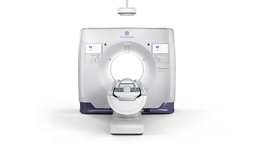
GE HealthCare has expanded its global clinical and research network across North America, Europe, and Asia to evaluate Photonova Spectra, a photon-counting computed tomography system. Following CE Mark, United States Food and Drug Administration 510(k) clearance, and regulatory approval from Japan’s Pharmaceuticals and Medical Devices Agency, seven academic medical centers are executing structured clinical trials to define imaging protocols, assess spectral biomarkers, and validate quantitative diagnostic workflows.
Diagnostic Imaging Constraints in Conventional Computed Tomography
Conventional energy-integrating computed tomography detectors convert incoming X-ray photons into visible light before generating an electrical signal, which introduces electronic noise, limits spatial resolution, and obscures low-energy spectral information. Overcoming these physical bottlenecks in medical imaging infrastructure requires direct photon-counting architectures capable of measuring individual X-ray photon energies simultaneously. Validating these capabilities across varied clinical disciplines—such as cardiology, pediatrics, and oncology—requires multi-center clinical benchmarking to standardize radiation dose parameters and reconstruction pipelines before widespread hospital deployment.
Detector Architecture and Technical Responsibilities
The technical evaluation pairs GE HealthCare’s hardware and reconstruction software with the clinical research infrastructure of seven partner institutions. GE HealthCare supplies the Photonova Spectra scanner, which utilizes proprietary Deep Silicon detector technology to capture individual photons directly. This semiconductor detector architecture delivers ultra-high definition spatial resolution alongside multi-energy spectral data within a single-scan workflow, enabling material decomposition and quantitative tissue characterization without separate dual-energy acquisitions.
GE HealthCare engineers, scientists, and product specialists collaborate directly with clinical investigators to refine image reconstruction algorithms, optimize spectral presentation workflows, and translate empirical performance metrics into future system updates.
Global Clinical Validation and Protocol Testing
The multi-center evaluation spans seven medical research facilities executing targeted technical assessments:
Keio University Hospital in Tokyo, Japan, investigates quantitative spectral imaging methods and emerging clinical applications.
Centro Cardiologico Monzino IRCCS in Milan, Italy, evaluates cardiovascular imaging workflows, quantitative myocardial assessment, and tissue characterization.
Universitätsspital Zürich in Zürich, Switzerland, tests coronary computed tomography angiography protocols through its Department of Nuclear Medicine, focusing on radiation dose reduction while maintaining diagnostic accuracy.
University of Wisconsin–Madison in Madison, Wisconsin, conducts technical image quality benchmarks and quantitative performance evaluations.
Stanford Medicine in Palo Alto, California, executes technical and human-subject studies covering image reconstruction methods, structural visualization, and spectral imaging biomarkers.
These programs expand upon post-CE Mark collaborations with UZ Brussel – Vrije Universiteit Brussel in Belgium for functional coronary analysis, and Rigshospitalet in Copenhagen, Denmark, which evaluates low-dose pediatric imaging and spectral characterization in complex clinical cases.
Professor Philipp Kaufmann, Director of the Department of Nuclear Medicine at Universitätsspital Zürich, stated the clinical objective: "We aim to determine how best to translate these capabilities into robust, dose-efficient CCTA protocols that support confident clinical decision-making."
Clinical Workflow and Diagnostic Impact
Consolidating ultra-high spatial resolution and quantitative spectral analysis into a single-scan workflow eliminates the need for repeat examinations and reduces cumulative patient radiation exposure. Standardized multi-center clinical data establishes validated protocols for material characterization, improves anatomical visualization in complex cardiovascular and pediatric cases, and accelerates the integration of quantitative biomarkers into routine diagnostic radiology.
Edited by Romila DSilva, Induportals Editor, with AI assistance.
www.gehealthcare.com

