
GE HealthCare Launches On-Device AI for Feeding Tube Detection on Mobile X-Ray Systems
GE HealthCare receives FDA 510(k) clearance for Critical Care Suite 3.0, embedding on-device artificial intelligence directly onto mobile X-ray systems to detect and localize enteric feeding tubes across adult and pediatric critical care workflows.
www.gehealthcare.com
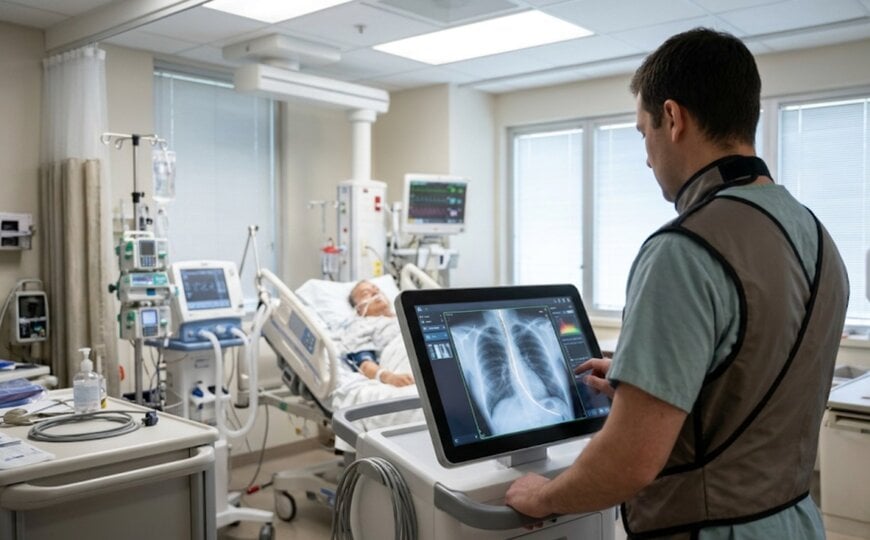
Emergency departments and intensive care units rely heavily on mobile radiographic imaging and enteral nutrition delivery, with mobile X-ray systems accounting for 75 percent of usage in emergency departments and 51 percent in intensive care units, while roughly 80 percent of intensive care patients receive nutrition via nasogastric or nasoenteric tubes. Verifying accurate enteric tube placement prior to initiating feeding is essential to prevent severe clinical complications such as aspiration pneumonia or pneumothorax. Traditionally, bedside clinicians must wait for acquired chest or abdominal radiographs to transmit over hospital networks to centralized Picture Archiving and Communication Systems (PACS) for remote radiologist review, introducing diagnostic delays and connectivity dependencies. Furthermore, extending automated tube-detection algorithms to pediatric patients introduces substantial anatomical and technical complexities due to smaller patient dimensions, narrower airway structures, and specialized low-dose imaging protocols. Resolving these point-of-care diagnostic bottlenecks requires edge-embedded computer vision models capable of localizing tube trajectories and anatomical landmarks directly on the mobile X-ray console without external cloud connectivity.
On-Device AI Architecture and Anatomical Localization
To deliver immediate point-of-care placement verification, GE HealthCare received 510(k) clearance from the United States Food and Drug Administration for Critical Care Suite 3.0. Unlike cloud-dependent diagnostic software, the algorithm suite executes directly on the mobile X-ray hardware, analyzing enteric tube tip and side-port locations immediately at the patient bedside without requiring internet access or external server routing.
The computer vision pipeline automatically detects enteric tubes, delineates the full tube path, and pinpoints the distal tip alongside the side port. Simultaneously, the software highlights the airways and diaphragm as structural reference points to assist clinicians in verifying whether the tube has traversed the esophagus into the stomach or inadvertently entered the bronchial tree. In clinical validation testing, Critical Care Suite 3.0 achieved 99.2 percent sensitivity for enteric tube detection and a 91.6 percent mean DICE similarity coefficient for localization accuracy against expert radiologist annotations.
Pediatric Expansion and Point-of-Care Workflow Integration
With version 3.0, Critical Care Suite extends its feeding tube assessment capabilities to pediatric patients for the first time, accommodating the distinct anatomical proportions and radiographic exposure profiles required in neonatal and pediatric intensive care settings. Larry Boyd, Vice President and General Manager of Global X-ray at GE HealthCare, stated that embedding intuitive AI into established mobile X-ray workflows brings actionable placement data closer to bedside clinicians across a broader patient demographic when rapid decisions are required.
Building on earlier generations of the Critical Care Suite portfolio—which introduced on-device algorithms for automated measurements, critical finding prioritization, and quality control—version 3.0 also incorporates Quality Care Suite software to flag image quality errors prior to PACS transmission. Adam Walbrun, Clinical Manager of Imaging Services at Emplify Health in La Crosse, Wisconsin, noted that immediate bedside access to feeding tube localization supports faster clinical decision-making and drives frequent provider requests for portable X-ray units equipped with the software.
Additional Context
This section details technical specifications and competitive benchmarking not included in the original product announcement.
In radiographic computer-aided detection and triage software governed by FDA Software as a Medical Device (SaMD) frameworks and IEC 62304 medical device software lifecycle standards, evaluating segmentation and localization performance relies on spatial overlap metrics such as the Dice similarity coefficient alongside sensitivity and specificity thresholds. Achieving a mean Dice score above 90 percent for narrow, low-contrast radiopaque catheter filaments superimposed over complex thoracic and abdominal anatomy requires deep convolutional neural network architectures trained on high-resolution digital flat-panel detector outputs. Executing inference directly on the mobile X-ray workstation's local graphics processing unit eliminates network latency, avoids Protected Health Information (PHI) transit risks outside the hospital firewall, and ensures continuous clinical availability during hospital network outages or disaster-response deployments.
Within the point-of-care radiographic AI sector, GE HealthCare’s on-device Critical Care Suite benchmarks against both embedded hardware-integrated suites and cloud-based radiology triage platforms, including Siemens Healthineers' AI-Rad Companion Chest X-ray, Philips' AI-enabled mobile radiography workflows (such as the MobileDiagnost wDR platform), Fujifilm's REiLI on-device AI suite, and specialized third-party radiology AI vendors such as Annalise.ai, Lunit (Lunit INSIGHT CXR), and Aidoc. While third-party radiology AI platforms typically ingest DICOM studies centrally after PACS routing to prioritize radiologist worklists across multiple scanner brands, embedding inference natively on the mobile X-ray acquisition console provides immediate visual overlays to the acquiring technologist and bedside intensivist before the portable unit leaves the patient's room. Expanding validated tube-tip and side-port segmentation to pediatric populations further differentiates the platform, as most commercial chest and abdominal AI algorithms remain restricted to adult cohorts due to the high anatomical variance across pediatric developmental stages.
Edited by Romila DSilva, Induportals Editor, with AI assistance.
www.gehealthcare.com

