
www.medical-devices.tech
11
'26
Written on Modified on
DePuy Synthes Launches VELYS Hip Navigation with AI
Next-generation software solution includes new AI capabilities to advance the standard of care in total hip arthroplasty (THA) by automating image interpretation, consistently identifying landmarks, and increasing procedural efficiencies.
www.jnjmedtech.com
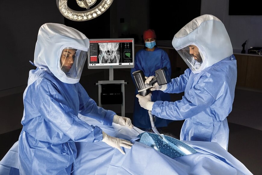
Use of VELYS™ Hip Navigation with AI Assistance demonstrated reproducible landmark identification and reduced workflow time by 57% compared to manual workflows (5.33 vs. 2.27 minutes)
DePuy Synthes has announced the U.S. commercial availability of VELYS™ Hip Navigation with AI Assistance, a next-generation surgical software platform engineered to automate image interpretation and anatomical landmark identification during total hip arthroplasty (THA).
Demographic Pressures and Operational Demands
Demographic projections indicate that total hip arthroplasty demand continues to scale in the United States, with approximately 850,000 annual procedures anticipated by 2030. This projection represents a 129% volume increase compared to statistical baselines from 2014.
While THA remains a highly successful orthopedic intervention, the expansion of patient volumes and concurrent operating room scheduling constraints have created a clear industrial requirement for digital surgery platforms. Hospitals and ambulatory environments increasingly require advanced systems capable of supporting streamlined workflows, predictable intraoperative execution, and rapid data extraction for real-time decision-making.
Algorithmic Architecture and Human-in-the-Loop Safeguards
The VELYS Hip Navigation system addresses these operational bottlenecks by utilizing a specialized artificial intelligence model to handle intraoperative imaging datasets. The software automates image interpretation to supply the surgical team with real-time, patient-specific biophysical metrics, including:
DePuy Synthes has announced the U.S. commercial availability of VELYS™ Hip Navigation with AI Assistance, a next-generation surgical software platform engineered to automate image interpretation and anatomical landmark identification during total hip arthroplasty (THA).
Demographic Pressures and Operational Demands
Demographic projections indicate that total hip arthroplasty demand continues to scale in the United States, with approximately 850,000 annual procedures anticipated by 2030. This projection represents a 129% volume increase compared to statistical baselines from 2014.
While THA remains a highly successful orthopedic intervention, the expansion of patient volumes and concurrent operating room scheduling constraints have created a clear industrial requirement for digital surgery platforms. Hospitals and ambulatory environments increasingly require advanced systems capable of supporting streamlined workflows, predictable intraoperative execution, and rapid data extraction for real-time decision-making.
Algorithmic Architecture and Human-in-the-Loop Safeguards
The VELYS Hip Navigation system addresses these operational bottlenecks by utilizing a specialized artificial intelligence model to handle intraoperative imaging datasets. The software automates image interpretation to supply the surgical team with real-time, patient-specific biophysical metrics, including:
- Changes in leg length
- Joint offset distances
- Acetabular cup placement positioning
The underlying AI algorithm was trained on geographically diverse, deidentified clinical datasets to establish broad categorical consistency. To ensure patient safety, the system operates under a strict "human-in-the-loop" structural paradigm. This configuration verifies that the attending surgeon retains complete manual control over final anatomical landmark placement, keeping clinical authority in human hands. By substituting manual image measurements with automated algorithmic analysis, the system accelerates data accessibility to enhance operating room decision-making and overall process efficiency.
Sharrolyn Josse, Global President of Joint Reconstruction at DePuy Synthes, stated that the platform represents a progressive step in the deployment of data-driven technologies aimed at improving surgeon choice and procedural consistency. Josse noted that the framework is engineered as a next-generation hardware and software configuration developed to limit procedural complexity while preserving full clinical command for the operating physician.
Integrated Hardware and Monitor Specifications
The AI-assisted navigation application runs on the VELYS Hub, an advanced, specialized hardware console designed specifically for data-intensive clinical care. The physical assembly includes a 32-inch 4K high-resolution monitor that offers a 79% larger active viewing zone than previous legacy display models.
To bridge the gap between imaging systems and computer screens, the hardware stack incorporates an integrated image-capture card that provides plug-and-play connectivity with standard surgical C-arms. Computational processing is driven by a GPU-optimized computer architecture, which provides the high-throughput mathematical rendering necessary to generate real-time intraoperative data screens during active surgery.
According to Christopher N. Johnson, D.O., an orthopedic surgeon at Ortho Northeast, establishing consistent component positioning and restoring patient-specific biomechanics represent the core foundations for long-term implant survival. Johnson added that digital navigation systems built with AI assistance have clear potential to minimize interoperative variability, optimize surgical workflows, and enhance intraoperative choice.
Additional Context
This section details technical specifications not included in the original news release.
Digital hip navigation systems provide an alternative to traditional mechanical alignment jigs, which are prone to manual visual alignment errors during component positioning. In total hip arthroplasty, the primary mechanical objectives are the restoration of the patient's anatomical center of rotation and the precise orientation of the acetabular cup component. The optimal orientation is typically defined by an anteversion angle of 15° ± 10° and an inclination (abduction) angle of 40° ± 10°, a spatial envelope historically referred to as Lewinnek's safe zone. Deviations from these target parameters drastically elevate the risk of post-operative joint dislocation, component impingement, eccentric polyethylene liner wear, and premature mechanical failure.
To extract these measurements intraoperatively without the sub-millisecond lag associated with legacy systems, modern digital surgery hubs leverage advanced computer vision algorithms, such as Convolutional Neural Networks (CNNs), running on dedicated graphics processing units (GPUs). The platform processes raw digital fluoroscopy images generated by standard mobile C-arm X-ray systems.
Upon receiving the DICOM image matrix via the integrated capture card, the AI model executes a multi-stage semantic segmentation pipeline. The network automatically filters out surgical instrumentation and implants to isolate osseous structures, accurately identifying critical anatomical landmarks such as:
Sharrolyn Josse, Global President of Joint Reconstruction at DePuy Synthes, stated that the platform represents a progressive step in the deployment of data-driven technologies aimed at improving surgeon choice and procedural consistency. Josse noted that the framework is engineered as a next-generation hardware and software configuration developed to limit procedural complexity while preserving full clinical command for the operating physician.
Integrated Hardware and Monitor Specifications
The AI-assisted navigation application runs on the VELYS Hub, an advanced, specialized hardware console designed specifically for data-intensive clinical care. The physical assembly includes a 32-inch 4K high-resolution monitor that offers a 79% larger active viewing zone than previous legacy display models.
To bridge the gap between imaging systems and computer screens, the hardware stack incorporates an integrated image-capture card that provides plug-and-play connectivity with standard surgical C-arms. Computational processing is driven by a GPU-optimized computer architecture, which provides the high-throughput mathematical rendering necessary to generate real-time intraoperative data screens during active surgery.
According to Christopher N. Johnson, D.O., an orthopedic surgeon at Ortho Northeast, establishing consistent component positioning and restoring patient-specific biomechanics represent the core foundations for long-term implant survival. Johnson added that digital navigation systems built with AI assistance have clear potential to minimize interoperative variability, optimize surgical workflows, and enhance intraoperative choice.
Additional Context
This section details technical specifications not included in the original news release.
Digital hip navigation systems provide an alternative to traditional mechanical alignment jigs, which are prone to manual visual alignment errors during component positioning. In total hip arthroplasty, the primary mechanical objectives are the restoration of the patient's anatomical center of rotation and the precise orientation of the acetabular cup component. The optimal orientation is typically defined by an anteversion angle of 15° ± 10° and an inclination (abduction) angle of 40° ± 10°, a spatial envelope historically referred to as Lewinnek's safe zone. Deviations from these target parameters drastically elevate the risk of post-operative joint dislocation, component impingement, eccentric polyethylene liner wear, and premature mechanical failure.
To extract these measurements intraoperatively without the sub-millisecond lag associated with legacy systems, modern digital surgery hubs leverage advanced computer vision algorithms, such as Convolutional Neural Networks (CNNs), running on dedicated graphics processing units (GPUs). The platform processes raw digital fluoroscopy images generated by standard mobile C-arm X-ray systems.
Upon receiving the DICOM image matrix via the integrated capture card, the AI model executes a multi-stage semantic segmentation pipeline. The network automatically filters out surgical instrumentation and implants to isolate osseous structures, accurately identifying critical anatomical landmarks such as:
- The tear-drop lines
- The bi-ischial line
- The medial femoral cortex
- The center of the femoral head
By calculating the precise spatial pixel transformations between pre-resection and intraoperative trials, the system computes differences in leg length and global femoral offset. Clinical data demonstrates that replacing manual landmark plotting with automated CNN tracking reduces overall intraoperative workflow times by 57% compared to traditional computer navigation techniques, lowering average image interpretation sequences from 5.33 minutes down to 2.27 minutes.
Edited by Romila DSilva, Induportals Editor, with AI assistance.
Edited by Romila DSilva, Induportals Editor, with AI assistance.

