
www.medical-devices.tech
11
'25
Written on Modified on
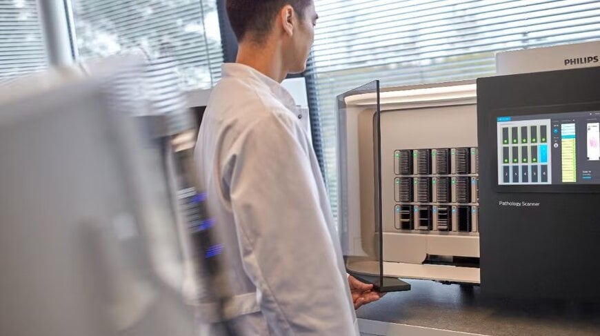
Philips Digital Pathology Scanner with DICOM Output
The Philips pathology scanner is the first in the world to offer native DICOM JPEG XL output, which reduces file sizes by up to 50% while maintaining high image quality.
www.philips.com
Pathology plays a crucial role in the diagnosis of a variety of diseases, particularly cancer, through examination of patient tissue samples. With an estimated 70% of important medical decisions involving laboratory or pathology tests, the availability of digitally stored pathology images is especially important as it has a significant impact on patient care. The digital transformation of pathology also means there is a growing need for scalable storage to meet new data volumes and computing resources, which also allows full AI adoption in clinical and research environments.
DICOM (Digital Imaging and Communications in Medicine) is the international standard for medical images and related patient information. Because digital pathology is a relatively new imaging modality compared to radiology and others, there was no established DICOM standard in place. As a result, vendors created their own proprietary formats.
Philips announced that it is expanding its SG300 and SG60 scanner offering with the Pathology Scanner SGi with configurable DICOM JPEG and DICOM JPEG XL output. As a result, it is the first in the world to offer native DICOM JPEG XL output. DICOM JPEG XL output files are up to 50% smaller while still providing the same high image quality, enabling pathology labs to store, manage, and analyze growing volumes of digital pathology data and enable more productive workflows in the cloud and on-premises.
“The adoption of DICOM in pathology marks an important shift toward achieving scalable, interoperable imaging workflows, said Imogen Fitt, Principal Analyst at Signify Research. “As pathology labs face mounting data volumes and storage demands, the move to standardized formats helps reduce infrastructure costs and enables integration with a wider range of AI tools. Philips’ support for DICOM JPEG and DICOM JPEG XL as a native output from its scanners illustrates how vendors are aligning with these trends, intending to support capabilities such as centralized archiving, cross-modality diagnostics, and remote collaboration for customers.”
In addition, the DICOM standard eases collaboration between pathology and other imaging modalities, scaling cross-modality integrated diagnostics and decision-making, as well as consolidation of IT infrastructure through the archiving of both radiology and pathology images in a single location and archive (a PACS or vendor-neutral archive).
www.philips.com
DICOM (Digital Imaging and Communications in Medicine) is the international standard for medical images and related patient information. Because digital pathology is a relatively new imaging modality compared to radiology and others, there was no established DICOM standard in place. As a result, vendors created their own proprietary formats.
Philips announced that it is expanding its SG300 and SG60 scanner offering with the Pathology Scanner SGi with configurable DICOM JPEG and DICOM JPEG XL output. As a result, it is the first in the world to offer native DICOM JPEG XL output. DICOM JPEG XL output files are up to 50% smaller while still providing the same high image quality, enabling pathology labs to store, manage, and analyze growing volumes of digital pathology data and enable more productive workflows in the cloud and on-premises.
“The adoption of DICOM in pathology marks an important shift toward achieving scalable, interoperable imaging workflows, said Imogen Fitt, Principal Analyst at Signify Research. “As pathology labs face mounting data volumes and storage demands, the move to standardized formats helps reduce infrastructure costs and enables integration with a wider range of AI tools. Philips’ support for DICOM JPEG and DICOM JPEG XL as a native output from its scanners illustrates how vendors are aligning with these trends, intending to support capabilities such as centralized archiving, cross-modality diagnostics, and remote collaboration for customers.”
In addition, the DICOM standard eases collaboration between pathology and other imaging modalities, scaling cross-modality integrated diagnostics and decision-making, as well as consolidation of IT infrastructure through the archiving of both radiology and pathology images in a single location and archive (a PACS or vendor-neutral archive).
www.philips.com

